Recently, the team led by Zhao Bing from the Institute of Organoid Research at Nanchang University and the Ministry of Education Organoid Resource Center achieved significant breakthroughs in the field of in vitro organoid culture for glands such as the meibomian gland and the adrenal gland. They published a series of research papers in the journals Protein & Cell and Stem Cell Reports, which were highlighted by Nature.
Organoid technology is a cutting-edge, disruptive technology in the biomedical field. It utilizes tissue stem cells to cultivate three-dimensional "micro-organs" in vitro that highly simulate the structure and function of real organs. The U.S. Food and Drug Administration (FDA) and the European pharmaceutical regulatory system have successively issued the Modernization Act 2.0 and various new methodology roadmaps, explicitly defining the important status of organoids in disease modeling and preclinical new drug development, thereby generally weakening the traditional paradigm where "animal experiments are the sole or mandatory path."
The Meibomian gland (MG) is a specialized sebaceous gland located in the eyelid. It secretes the outer lipid layer of the tear film through a holocrine secretion mode, which is crucial for reducing tear evaporation and maintaining ocular surface homeostasis. Under physiological conditions, MG acinar basal cells are capable of self-renewal and differentiation into mature meibocytes; the latter synthesize lipids during their migration toward the center of the acinus and eventually rupture to release their lipid content.
Meibomian gland dysfunction (MGD) is the primary cause of dry eye disease. The global prevalence of dry eye disease is as high as 38%-68%, characterized by symptoms including vision decline, ocular stinging, and ocular surface inflammation. Currently, interventions are limited to frequent supplementation with artificial tears, and there are no direct drugs available to salvage meibomian gland function. The challenge in drug development is largely attributed to the lack of disease models, leading to unclear pathogenic mechanisms and difficulties in intervention research.
Zhao Bing’s team achieved a significant breakthrough in research on meibomian gland organoids and dry eye disease models. They published a research article in Protein & Cell titled “Human meibomian gland organoids to study epithelial homeostasis and dysfunction.” They constructed, de novo, mouse and human meibomian gland organoids capable of long-term stable expansion, fully recapitulating the meibomian gland cell lineage and lipid secretion function in vitro.

The meibomian gland organoids accurately simulate responses to potential dry eye-inducing drugs and intervention drugs. Furthermore, after in situ transplantation, they can reconstruct human meibomian gland tissue and lipid secretion function within experimental animals. Single-cell expression profiling and lipidomics analyses performed in the organoids elucidated key regulatory signals for meibomian gland homeostasis maintenance and disease occurrence. Importantly, the team also utilized the organoids to confirm the significant improvement effect of the FGF10 cytokine drug on meibomian gland dysfunction, providing important support for its efficacy and pharmacological research.

The meibomian gland organoids and dry eye disease models constructed in this study provide important model tools for delving into meibomian gland physiological and pathological mechanisms, accelerating drug development in the field of dry eye disease, and exploring meibomian gland regenerative medicine.

Concurrently, the team published a research article in Stem Cell Reports titled “Human adrenocortical organoids for tissue regeneration and disease modeling.” They constructed, de novo, human adrenocortical organoids and Cushing’s syndrome (adrenocortical adenoma) disease models. The human adrenocortical organoids can respond to physiological stimuli to secrete glucocorticoids in vitro, and regenerate human adrenocortical tissue after subcutaneous transplantation, enabling the rescue of mice that have had their adrenal glands removed.
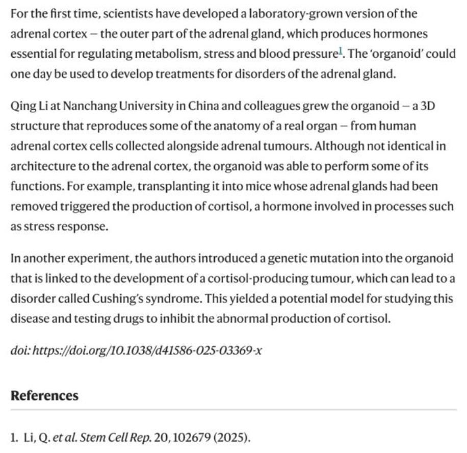
Following the publication of this work, it was highlighted by Nature magazine. In an article titled “Gland-in-a-dish secretes stress hormone like the real thing,” it was noted: “Scientists have grown the adrenal cortex in the lab for the first time. This is the outermost layer of the adrenal gland, which produces essential hormones that regulate metabolism, handle stress and modulate blood pressure. One day, such organoids might be used to treat adrenal disease... Nanchang University used human adrenocortical cells collected from near adrenal tumors to cultivate adrenocortical organoids. The findings are a model with huge potential that can be used to study this disease and test new drugs to suppress abnormal cortisol secretion.”
Nanchang University is the first corresponding unit for both works, and patent protection and industrial transformation have been realized through horizontal agreements. Director Hong Jiaxu of the Eye & ENT Hospital of Fudan University, Director Jiang Jingjing of Zhongshan Hospital Fudan University, and Bozhen Biology provided important collaborative support.
The Institute of Organoid Research at Nanchang University is the only secondary research institution named after organoids among China’s "211 Project" universities; the Ministry of Education Organoid Resource Center is the first ministerial-level systematic resource platform for organoids. The Institute and the Resource Center emphasize the construction of talent teams and continuously recruit graduate students, postdoctoral fellows, and young teachers interested in organoid research, technology translation, and interdisciplinary fields such as AI to join them, in order to promote the vigorous development of organoid research at Nanchang University.
Original Articles:https://doi.org/10.1093/procel/pwaf095
https://doi.org/10.1016/j.stemcr.2025.102679
https://doi.org/10.1038/d41586-025-03369-x

 Address: No. 1299 Xuefu Avenue, Honggutan District, Nanchang City, Jiangxi Province
 Tel:+86-0791-83968485
 E-mail:ibi@ncu.edu.cn
 NANCHANG UNIVERSITY

